Learn About the New Optomap
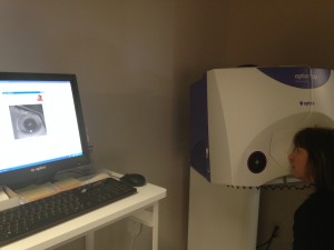
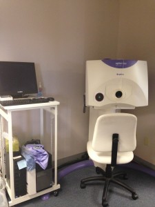 In 2012, at Sharonville Evendale Eyecare we introduced a new way for Dr. Murray to view your retina, the Optomap Imaging System, some exciting new technology that allows the doctor to take a picture of the retina and see a wider angle than previously possible. Although many current patients have already experienced the Optomap, it’s always valuable to be reeducated about it; and for those new patients who are wondering what in the world we’re talking about with all this Optomap stuff, now’s the chance to find out!
The Medical Science Behind the Optomap
Located in the back of your eye, your retina is the only place in your body where blood vessels can be seen directly. Because of this fact, diseases like stroke, hypertension, diabetes and heart disease can be seen in the retina. While you might not notice any of the symptoms associated with these diseases and conditions, their signs may still be present in your retina.
Optomap vs. Traditional Imaging Methods
The Optomap is unique in that it is able to capture 80% of your retina in one single panoramic image, whereas traditional imaging methods only show up to 15% of the retina at a time.
The Optos website has a great visual comparison in which you can toggle between images to see for yourself what traditional imaging can show vs. what the Optomap is able to present to your eye doctor.
Benefits of the Optomap
The benefits of the Optomap should be clear. Early detection of retinal diseases and other health conditions can be spotted well in advance of their onset. The Optomap can provide early protection from vision impairment and blindness, as well as detect early on life threatening diseases like cancer, stroke and cardiovascular disease.
Questions About the Optomap
The Optos website provides a great question-and-answer section about the Optomap. Here is a sample of some common ones we’ve received:
Is an optomap safe for children?
Yes. In fact, many vision problems begin in early childhood, so it’s important for children to receive quality routine eye care.
Does it hurt?
No. It is completely comfortable and the scan takes less than a second.
How often should I have an optomap?
This is a decision that should be made by your doctor. However, it is generally recommended that you have an optomap each time you have an eye exam.